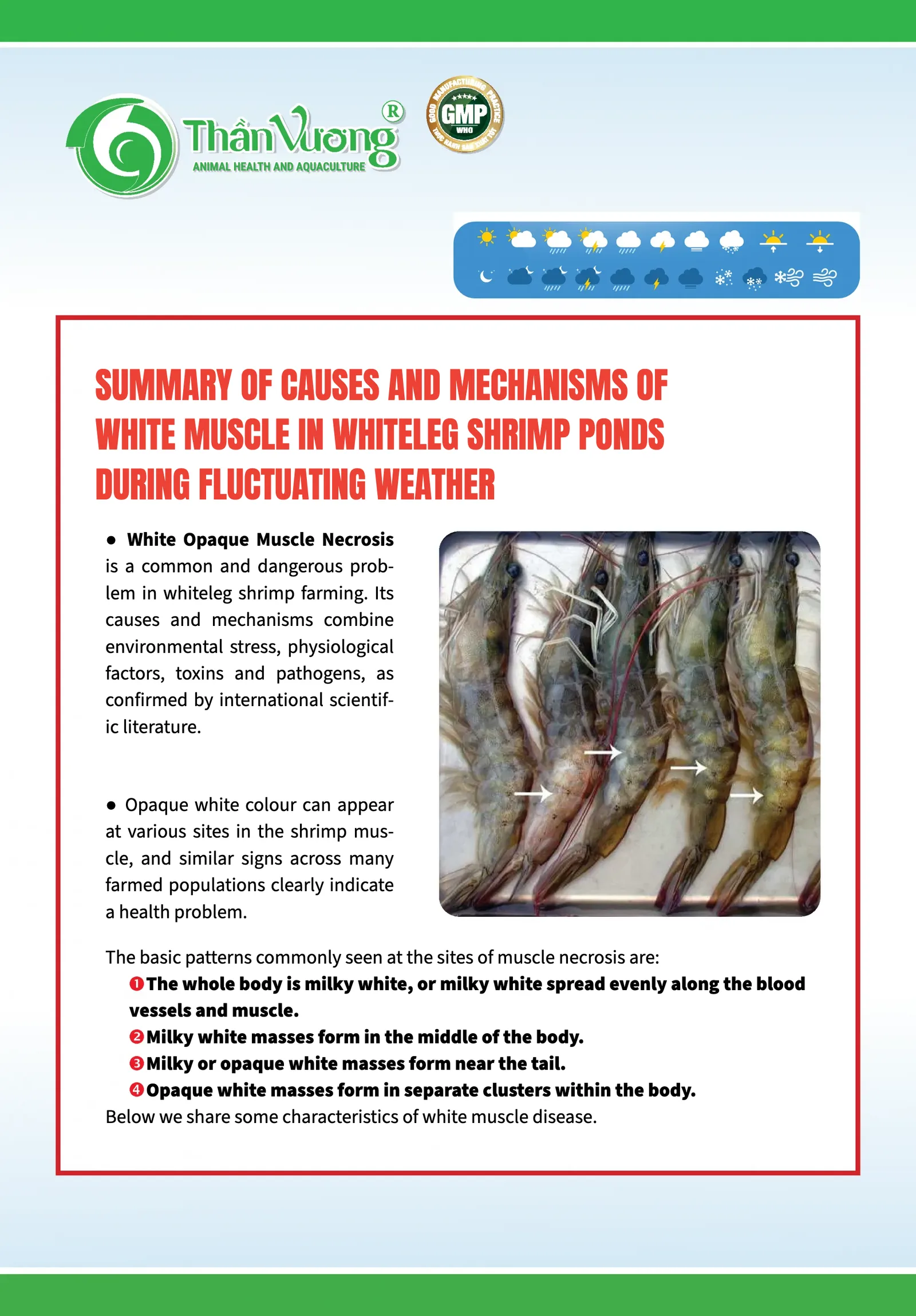
- White Opaque Muscle Necrosis is a common and dangerous problem in whiteleg shrimp farming. Its pathogenesis, causes and mechanisms involve a combination of environmental stress, physiological factors, toxins and pathogens, as confirmed by international scientific literature.
- Opaque white colour can show up in different parts of the shrimp body, but similar patterns across many farmed populations are a clear sign that the shrimp have health problems.
The basic patterns usually seen where muscle necrosis occurs are:
- The whole body is cloudy white, or cloudy white but spread evenly along the blood vessels and muscle.
- Cloudy white patches form in the middle of the body.
- Cloudy white or opaque white patches form near the tail.
- Opaque white patches form in separate clusters in the body.
Below we share some characteristics of white muscle disease.
1. Hypoxia stress and environmental toxins
Photo in the original: external appearance of naturally affected shrimp. (a) White-red patches on the epidermis (arrows). (b) White patches spread across the epidermis, including the abdominal segments (arrows). Source: Intriago et al., 2024.
- Changes in haemocyanin structure: hypoxia stress, toxins, mineral deficiency (copper) and pH swings drive conversion between the forms of haemocyanin: oxy-haemocyanin (Cu2+), deoxy-haemocyanin (Cu1+) and met-haemocyanin (Cu2+ oxidised, no longer able to carry oxygen). This limits the oxygen reaching muscle cells, causing cell death, oedema, protein coagulation and opaque white tissue damage (He et al., 2024).
- When oxygen levels in the shrimp's blood and muscle fall, the muscle gradually turns from its natural translucent colour to cloudy white or opaque white. This rarely happens in ponds with dissolved oxygen above 4 mg/l. However, when shrimp are overexerted, for example when they run in schools, or when farmers net shrimp to thin or transfer them and the shrimp flick, jump and swim too hard, oxygen in the blood cannot be delivered fast enough and the muscle turns cloudy white. In experiments, when dissolved oxygen was lowered below 1 mg/l, scientists saw the shrimp muscle gradually turn opaque across the whole body.
- During the crop, at certain times, such as after feeding when the shrimp's oxygen demand rises sharply, or when the algae crash or heavy rain causes falling oxygen, rising toxic gases and changes in pH and temperature, the shrimp become exhausted and carry oxygen poorly, which also makes them prone to cloudy muscle. In many grow-out ponds, farmers measure consistently high dissolved oxygen, but that does not prove the shrimp can take up enough oxygen to meet their needs, because toxic gases such as NO2 and NH3, and turbidity, are far too high at spots such as the pond centre. NH3 disrupts the immune system (Luo et al., 2024), damages the gills and blocks the shrimp's metabolism, so it often causes acute toxicity. NO2 disrupts and affects activity inside the cells; it acts more slowly, but it impairs oxygen transport and nutrient absorption. Weak shrimp then go through incomplete moulting and are lost here and there. A warning sign: shrimp that easily show cloudy muscle when the feeding trays are lifted may be stressed or in poor health.
Figure in the original: hepatopancreas ruptured by environmental stress. Source: Mobaraki et al., 2022.
Under stress from the environment or from nutritional deficiency, such as minerals and amino acids, shrimp are prone to muscle cramps and have difficulty straightening the body, similar to cramps during exercise. These muscles do not get oxygen in time, which then leads to muscle necrosis. Two situations are common. In juvenile shrimp, cloudy dorsal muscle (or a "broken back") often appears from a strong sideways flicking reflex when startled, or the shrimp moult while short of oxygen, so the muscle cramps and they drop to the bottom. In large shrimp, the heavy shell makes them flick backwards and moult more slowly, so broken back and cloudy dorsal muscle become less common; however, muscle necrosis from incomplete moulting / stuck shell increases a lot, because they carry a large, heavy shell and need more tail force to get out, and this overexertion makes them prone to cloudy tail muscle.
In shrimp farming, two other forms of cloudy tail muscle are also seen: those caused by bacteria and those caused by viruses.
2. Environmental shock, minerals and pH
- Sudden changes: sudden changes in temperature, salinity or pH, or a shortage of the minerals Ca, Mg, K or Cu, upset the ion balance, causing weak muscle contraction, oedema and necrosis.
- Abnormal pH: abnormal pH (<7 or >8.5) and alkalinity that is too high or too low inhibit enzymes and increase oxidative reactions, leading to necrosis of muscle tissue.
3. Infection by biological agents: viruses, bacteria, microsporidia
Viruses, bacteria, fungi and microsporidia are, broadly, all foreign agents that invade the shrimp's body. Lacking an adaptive immune system, shrimp rely on their effective innate cellular and humoral immune responses to fight invading bacteria. At the site of infection, shrimp tissue may turn opaque because of bacterial invasion, damage from oxidative reactions (H2O2) and clotting reactions that try to stop the pathogen from spreading and invading the body. The shrimp muscle then often turns opaque in particular places, like cotton wool, or opaque white along the blood-vessel tissue that supplies the body and around the hindgut, and gradually spreads.
Viral muscle necrosis: the viruses IMNV and PvNV are proven causes in pond-farmed whiteleg shrimp. The virus attacks muscle tissue, creating multifocal necrotic areas on histopathology, inflammation and death of muscle fibre cells, so the muscle looks opaque white. Photo source in the original: Jose et al.
Diagram in the original: model of IMNV entry (Rong et al. 2025), covering viral entry and expression, metabolic reprogramming (carbohydrate, amino acid and lipid metabolism), autophagy, ER stress and host-environment interaction, leading to muscle necrosis.
- Infectious myonecrosis (IMN) is caused by infection with infectious myonecrosis virus (IMNV), a totivirus. The main target tissue of IMNV is striated muscle. Shrimp infected with IMNV have focal or widespread white necrotic areas in the striated muscle, especially in the distal abdominal segments and the tail fan.
- Penaeid white tail disease (PWTD) in whiteleg shrimp is caused by a virus called Penaeus nodavirus, or PvNV. Muscle necrosis in shrimp often leads to opaque white lesions in the tail muscle, in response to environmental factors such as low dissolved oxygen, sudden changes in temperature or salinity, or other stress.
- Bacteria (Vibrio harveyi, Bacillus cereus): bacterial enzymes and toxins lyse muscle cell membranes, causing muscle breakdown and multifocal necrosis, with the common sign of opaque white muscle.
- IMNV infects the host shrimp's muscle cells, leading to cell necrosis. The virus does this by suppressing the host's innate immune response, which lets it invade and spread. It then takes over host cell processes, including inducing oxidative stress, remodelling the cytoskeleton and activating cell-death pathways such as apoptosis and ferroptosis, which in the end leads to widespread death of muscle cells.
- The microsporidia behind cotton shrimp disease include one or more microsporidian genera (Pleistophora, Thelohania, Perezia, Agmasoma and Ameson). They attack shrimp muscle and cause necrosis, forming white, cotton-ball-like inflammatory foci. These are the result of inflammation, muscle destruction, immune clotting reactions and the phagocyte response trying to fight the superinfection. Photo source in the original: Robin et al. 2022.
- Perezia spp. destroy muscle and gradually replace it with masses of developing parasite stages and spores. In the late stage of infection, the parasite can invade other tissues and organs in the same shrimp, such as heart muscle cells, epithelial cells and the connective tissue of the antennal gland.
- Agmasoma penaei affects the gonads, the heart and blood vessels, the gills, the hepatopancreas and the midgut, producing enlarged white gonads or many swollen white lumps. Microsporidia in shrimp can cause serious outbreaks in farmed populations, but the disease is usually rare because it needs a fish intermediate host to complete its life cycle (Roger, 2022).
4. Heavy rain and seasonal change
Heavy rain lowers water quality and causes mass algae die-off, a build-up of toxic gases and organic matter, and bacterial blooms. The combined stress raises the risk of white muscle disease and increases mortality.
References
- Intriago, P., Montiel, B., Valarezo, M., Arteaga, K., Romero, X., & Montenegro, A. (2024). Bacterial muscle necrosis in cultured Penaeus vannamei in Latin America. Aquaculture Research and Engineering, 2024, Article 9919229. https://doi.org/10.1155/are/9919229
- Chuchird, N., Chongprachavat, N., Suanploy, W., Kitsanayanyong, L., Phansawat, P., Keetanon, A., Wimanhaemin, P., & Rairat, T. (2024). Investigation of pale shrimp disease in Pacific white shrimp (Litopenaeus vannamei) caused by Photobacterium damselae subsp. damselae in low salinity culture conditions. Aquaculture Reports, 39, 102416. https://doi.org/10.1016/j.aqrep.2024.102416
- Melena, J., Tomalá, J., Panchana, F., Betancourt, I., Gonzabay, C., Sonnenholzner, S., Amano, Y., & Bonami, J.-R. (2012). Infectious muscle necrosis etiology in the Pacific White Shrimp (Penaeus vannamei) cultured in Ecuador. CENAIM-ESPOL Technical Report. Guayaquil, Ecuador.
- Alolod, G. A. L., Matsumoto, S., Koiwai, K., Kondo, H., & Hirono, I. (2024). First report of white muscle disease caused by Photobacterium damselae subsp. damselae in Kuruma shrimp (Penaeus japonicus). Aquaculture, 593, 741278. https://doi.org/10.1016/j.aquaculture.2024.741278
- Jithendran, K. P., Krishnan, A. N., Jagadeesan, V., Anandaraja, R., et al. (2021). Co-infection of infectious myonecrosis virus and Enterocytozoon hepatopenaei in Penaeus vannamei farms in the east coast of India. Aquaculture Research, 52(8), 3593-3602. https://doi.org/10.1111/are.15304
- He, H., Huang, S., Geng, N., Weng, S., He, J., & Li, C. (2024). Acute hypoxia stress mediates HIF-1α-Yki-Cactus axis to facilitate the infection of Vibrio parahaemolyticus in Litopenaeus vannamei. Frontiers in Immunology, 15, 1476309. https://doi.org/10.3389/fimmu.2024.1476309
- Luo, Z., Zhou, F., Jiang, S., Huang, J., Yang, L., Yang, Q., Shi, J., Li, E., Ma, Z., & Li, Y. (2024). Immune and physiological responses in Penaeus monodon to ammonia-N stress: a multi-omics approach. Frontiers in Immunology, 15, 1520887. https://doi.org/10.3389/fimmu.2024.1520887
- Mobaraki, S., Javadzadeh, N., Mabudi, H., Hafezieh, M., & Khodadadi, M. (2022). The effect of dietary fatty acids on the incidence of the redhead syndrome in white leg shrimp (Litopenaeus vannamei) under environmental stress. Iranian Journal of Fisheries Sciences. https://doi.org/10.22092/ijfs.2022.127398
- Behera, P. C. (2022, February 2). Emerging skeletal muscle necrosis diseases in Vannamei shrimp (Litopenaeus vannamei). Engormix. Retrieved from https://en.engormix.com/aquaculture/aquaculture-health/emerging-skeletal-muscle-necrosis_a48501/
- Rong, Y., Qiao, Y., Cao, X., Jiang, G., Cheng, J., Jiang, H., & Shen, H. (2025). Integrated transcriptomic and metabolomic analysis reveals molecular mechanisms of infectious myonecrosis virus (IMNV) pathogenesis in Litopenaeus vannamei muscle tissue. Aquaculture, 604, 742443. https://doi.org/10.1016/j.aquaculture.2025.742443
- Overstreet, R. M., & Hawkins, W. E. (2017). Diseases and mortalities of fishes and other animals in the Gulf of Mexico. In C. H. Ward (Ed.), Habitats and biota of the Gulf of Mexico: Before the Deepwater Horizon oil spill (pp. 1589–1738). Springer. https://doi.org/10.1007/978-1-4939-3456-0_6
- Chong, R. S.-M. (2022). Cotton shrimp. In F. Kibenge, B. Baldisserotto, & R.-S. Chong (Eds.), Aquaculture pathophysiology: Volume II. Crustacean and mollusks diseases (pp. 249–251). Academic Press. https://doi.org/10.1016/B978-0-323-95434-1.00036-X
- Han, J. E., Lee, S. C., Park, S. C., & Le Groumellec, M. (2019, September 9). The microsporidium Perezia sp. and cotton shrimp disease: Detection by histopathology, and new in situ hybridization and PCR methods. Responsible Seafood Advocate. https://www.globalseafood.org/advocate/the-microsporidium-perezia-sp-and-cotton-shrimp-disease/
English text adapted from Than Vuong's original Vietnamese infographic.