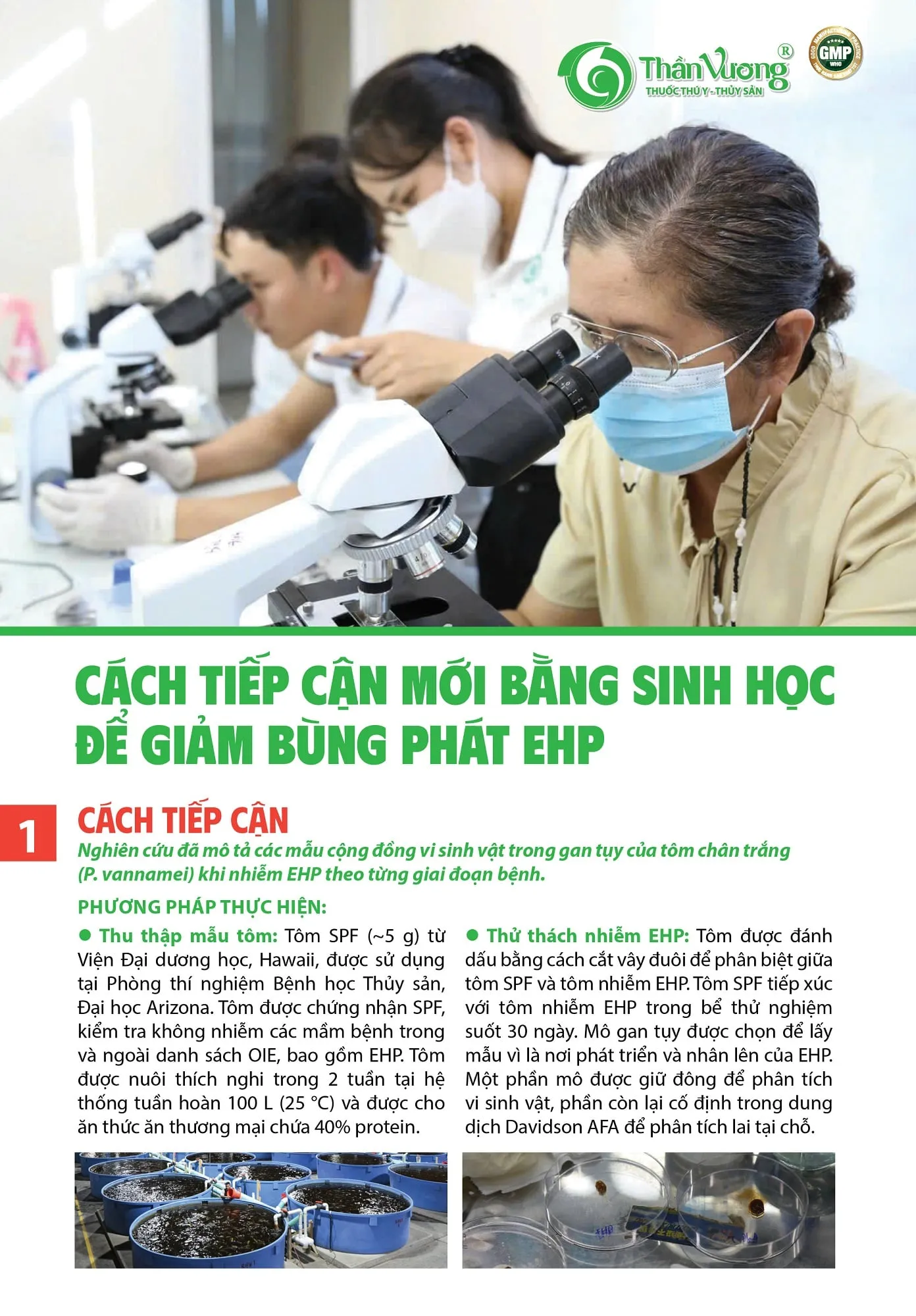
1. The approach
The study described the patterns of microbial communities in the hepatopancreas of whiteleg shrimp (P. vannamei) infected with EHP, stage by stage through the disease.
Methods
- Shrimp samples: SPF shrimp (~5 g) from the Oceanic Institute, Hawaii, were used at the Aquaculture Pathology Laboratory, University of Arizona. The shrimp were certified SPF and tested free of pathogens on and off the OIE list, including EHP. They were acclimated for 2 weeks in a 100 L recirculating system (25 °C) and fed a commercial feed containing 40% protein.
- EHP challenge: Shrimp were marked by clipping the tail fin to tell SPF shrimp apart from EHP-infected shrimp. SPF shrimp were kept together with EHP-infected shrimp in test tanks for 30 days. Hepatopancreas tissue was chosen for sampling because it is where EHP develops and multiplies. Part of the tissue was frozen for microbial analysis; the rest was fixed in Davidson's AFA solution for in situ hybridization.
The cyclical pattern of EHP infection
The study recorded a cyclical pattern of EHP infection in P. vannamei. The early stage (first 12 days) showed beneficial microbes that support nutrient absorption, while EHP was detected in only a few individuals. The development stage (15–24 days) saw EHP intensify, beneficial microbes decline and opportunistic bacteria and fungi increase. The late stage (27–30 days) showed beneficial microbes completely replaced by pathogenic microbes.
| Indicator | Healthy state (early stage) | Dysbiosis (late stage) |
|---|---|---|
| Beneficial bacteria and fungi | High | Low |
| Pathogenic bacteria and fungi | Low | High |
| Bacterial diversity | High | Decreased |
| Fungal diversity | Low | High |
| Nutrient absorption | High | |
| Energy reserves | Decreased | |
| Infection process | Increased |
Between the two states lies a transition stage. Along the bottom, the diagram shows the EHP infection level rising and the host environment changing.
Figure 1. Modulation of the hepatopancreas microbiome in EHP-infected shrimp (P. vannamei). The schematic shows the main changes in the host's hepatopancreas environment during EHP infection (30 days), by the disease stages recorded in this study. Infection intensity reflects the cyclical pattern of EHP: larger EHP shapes indicate higher infection levels and smaller EHP shapes indicate lower levels. Blue bacteria: beneficial microbes; grey dots: EHP; red bacteria: opportunistic and pathogenic microbes. Created with BioRender (http://biorender.com/).
Microbial composition
- Bacteria: A total of 1,895 bacterial OTUs were identified, mainly in the phyla Proteobacteria, Firmicutes and Tenericutes. Genera such as Pseudomonas, Comamonas and Agrobacterium were associated with the early stage. Vibrio and Aquimarina appeared clearly in the late stage.
- Fungi: A total of 213 fungal OTUs were identified, with the phyla Basidiomycota and Ascomycota the most common. The genus Naganishia appeared mainly in the development stage, and Malassezia dominated the late stage, linked to microbial disruption.
Changes in microbial function
- Early stage: the microbiome supports digestion and nutrient absorption, such as fatty acid synthesis and membrane transport.
- Development stage: ribosome synthesis (a ribosome is a small organelle inside the cell whose main job is to make protein) begins to emerge, along with a rise in opportunistic microbes.
- Late stage: microbial functions shift to pathways linked to the growth of pathogenic bacteria such as Vibrio cholerae, and to carbohydrate-degrading activity in fungi.
Discussion
The results show that EHP can cause a chronic, cyclical pattern of infection, in which infection intensity changes over time and never disappears completely. This is consistent with reports of chronic diseases in other species. Dysbiosis (imbalance of the microbiome) was recorded: beneficial microbes fell in density or were replaced, while opportunistic bacteria and fungi grew strongly.
Applications and recommendations
These findings suggest that beneficial microbes such as Pediococcus acidilactici, S. boulardii, Bacteroides and Pseudomonas can support the development of probiotic products to prevent and control EHP infection. The study also suggests that the microbiome could be adjusted to improve shrimp health and disease resistance.
Conclusion
This study gives a comprehensive view of how the microbiome in the hepatopancreas of EHP-infected shrimp is modulated, showing a cyclical pattern of disease development. The shift in microbial composition and function from healthy to diseased was clearly recorded at each disease stage, opening new directions for the control and prevention of hepatopancreatic microsporidiosis in shrimp.
2. How EHP infects shrimp
The EHP life cycle is fairly simple, with only 2 forms: spores and meronts (the stage that develops and multiplies inside the host). Once inside the shrimp, EHP infects the hepatopancreas: the spore extrudes its polar tube, pierces the membrane of a hepatopancreatic tubule cell, injects its genetic material into the cell, replicates and forms many new spores. These burst the cell, are released and go on to infect other cells. Within 1 week, 1 spore can produce up to 1,000 spores.
The diagram shows a mature EHP spore infecting a shrimp hepatopancreatic tubule cell. The result is reduced growth, metabolism and immunity.
The hepatopancreas cells are destroyed and gradually slough off, seriously impairing hepatopancreas function. In addition, during infection shrimp are more easily attacked by opportunistic pathogens such as Vibrio bacteria or viruses.
3. Methods for detecting EHP
As noted above, the clinical signs of EHP infection in shrimp are not specific, so EHP cannot be diagnosed at the pond. The following methods are currently available to detect EHP in shrimp:
| Method | Notes |
|---|---|
| Fresh mount of hepatopancreas tissue under the microscope, 100x objective or higher | Easy to do and costs nothing. |
| Histopathology of hepatopancreas samples | More accurate, but harder to do and needs many different chemicals and stains. |
| Molecular methods: PCR, nested PCR or real-time PCR | Very reliable, but expensive. |
4. Tackling the EHP problem
How FLASH helps reduce Enterocytozoon hepatopenaei (EHP)
The EHP-reduction process with FLASH:
- Step 1: Build protection. FLASH helps protect the hepatopancreatic tubule cells, helping reduce the risk of EHP getting in.
- Step 2: Help keep EHP in check. FLASH helps keep opportunistic bacteria under control and helps limit the development and spread of EHP spores.
Result: lasting shrimp health. Lower risk of EHP infection, a protected hepatopancreas and steady shrimp growth. FLASH: optimal protection for a successful shrimp crop.
Preventing EHP
1. Manage the culture environment
- Maintain good water quality: keep culture water of high quality, exchange water regularly and check parameters such as temperature, pH and salinity. This helps reduce stress on the shrimp and limits the conditions for EHP to develop.
- Control stocking density: avoid stocking too densely, as this can raise the risk of disease spreading and lower water quality.
2. Use disease-free seed
- Only use seed that has been tested and certified free of EHP and other diseases. This limits the source of infection from the start.
3. Strengthen beneficial microbes
- Add probiotic products containing beneficial bacteria such as Pediococcus acidilactici, S. boulardii, Bacteroides and Pseudomonas to improve the gut and hepatopancreas microbiome, support digestion and help strengthen disease resistance.
4. Sound nutrition
- Provide high-quality, nutrient-rich feed that is free of bacterial contamination to keep shrimp in the best health. Clean, nutritionally complete feed helps strengthen the shrimp's natural resistance.
5. Biosecurity practices
- Disinfect and clean equipment: regularly clean and disinfect ponds, tools and related equipment to stop pathogens from spreading.
- Control the water source: make sure incoming water is free of pathogens, and limit the entry of organisms that may carry pathogens into the pond.
Pond preparation:
- Dry the pond for 5–7 days to destroy EHP pathogens.
- For ponds that have had EHP before, clean thoroughly and dry the pond for 10–15 days before the new crop. Spray NaOH 3% or KOH 1% solution over the pond surface, or apply quicklime (CaO) at 500–600 kg/1,000 m2, to raise the pH (above 12) and trigger EHP spores to germinate. Spores that germinate with no host die quickly and can no longer infect.
- Soak tools and equipment from the old crop in the solutions above for 12 hours.
Incoming water preparation:
- Incoming water is settled and filtered through several settling ponds, with a long settling time.
- At the end of the settling system, water is dosed with potassium permanganate (KMnO4) at 6 ppm.
- Water moved into the ready (reservoir) ponds is disinfected with chlorine at 35–40 ppm and left for a long time, 7–10 days, before being supplied to the culture ponds.
A typical farm model: water settles and is filtered through several ponds before it reaches the ready ponds. It is disinfected with chlorine and stored alternately in 2 ready ponds to supply the culture ponds.
6. Monitoring and early detection
- Test regularly with methods such as PCR and in situ hybridization (ISH) to detect EHP early. Early detection allows timely intervention and adjustment of the culture environment, preventing disease outbreaks.
RAPID quick test kit
- Fast: on-site testing, with results in just 50 minutes.
- High accuracy: agreement with laboratory testing of > 93%*, giving accurate results.
- Easy to use: a simple test procedure with no complex equipment; it can be done right on site.
- Cost saving: more than 30 times cheaper than sending samples to a lab.
Detect EHP early at low cost.
Conclusion
EHP is a major challenge for shrimp farming because it spreads quickly and seriously weakens shrimp health. Understanding the causes of the disease and applying suitable preventive measures are essential to protect shrimp health and productivity. Measures such as environmental management, disease-free seed, beneficial microbes, sound nutrition and continuous monitoring help reduce the risk of disease and protect the success of shrimp farming.
5. Using FLASH as an in-feed product
What FLASH does
- Helps regulate quorum sensing without affecting beneficial bacteria.
- Helps strongly limit the multiplication of harmful bacteria and helps reduce toxin production and biofilm formation.
- Helps reduce EHP outbreaks.
- Helps reduce how often antibiotics are used, putting a biological product first: FLASH, to help limit EHP development.
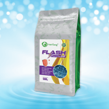
FLASH (500 g pack) helps reduce EHP outbreaks driven by the growth of opportunistic bacteria in the hepatopancreas.
Results: hepatopancreas and gut observations
| Observation | FLASH, DOC 57 | Control, DOC 57 |
|---|---|---|
| Lipid droplets in the tubules | 80–100% lipid droplets in the tubules | Mostly empty lipid droplets in the tubules |
| Tubule surface | Still normal | Starting to become jagged / wrinkled |
Shrimp at 32 days: no pale hepatopancreas and no weak gut.
Comparison of shrimp hepatopancreas and gut when using FLASH against the control.
How to use
| Stage | Dose | Frequency / duration |
|---|---|---|
| Preventive dose | ||
| Early stage and high-risk growth stage | 3–5 g FLASH/kg feed | 2 feedings/day (morning, afternoon) |
| Weight-gain stage | 3 g FLASH/kg feed | |
| Corrective / booster dose | ||
| Shrimp showing signs of EHP infection | 5–10 g FLASH/kg feed | 10–15 days |
Note: in rainy weather, depending on how much rain falls, add lime, minerals and anti-stress products to stabilise the environment quickly. Rain is the fastest driver of rising EHP load.
Benefits
- Helps reduce pathogen entry.
- Shrimp grow fast, with less stress.
- Low cost: the low dose does not harm beneficial bacteria in the shrimp gut.
- Fewer health problems and faster growth because fewer antibiotics are used.
Stability and key features
FLASH is an integrated solution that helps strengthen gut health and supports immunity in shrimp and fish. Below is a summary of its main features in terms of stability and effectiveness:
- Selective antibacterial action: helps reduce pathogenic bacteria while supporting beneficial bacteria, maintaining microbial balance.
- Quorum sensing inhibition: helps block communication between bacteria, reducing virulence and limiting biofilm formation.
- Gut microbiota diversity enhancement: improves the gut microbiome and supports digestion and nutrient absorption.
- Antiparasitic activity: helps control microsporidia and supports hepatopancreas health.
- Immunomodulation: helps strengthen natural immunity so the animals resist disease better.
English text adapted from Than Vuong's original Vietnamese infographic.